- Categories :
- More
Medical Imaging: Are We Getting Too Much Radiation?
By:
 The Woodlands Institute for Health & Wellness - Mila McManus MD
| Published 11/18/2026


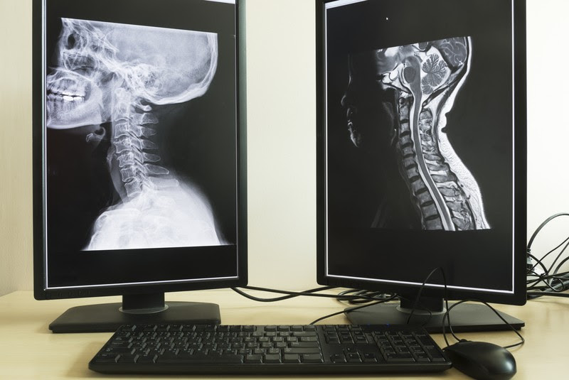
CT scans, X-rays, mammograms, and other imaging tests can provide valuable information about our health. But how much exposure risk is too much, and what should we know before scheduling another scan?
Medical imaging has transformed healthcare, helping physicians detect disease, evaluate injuries, and guide treatment. But the technologies used to create these images are not all the same. Imaging that uses ionizing radiation—particularly CT scans and some nuclear medicine procedures—deserve special attention because exposure can damage cellular DNA and, depending on the dose and circumstances, increase cancer risk. Understanding which tests involve radiation, how exposure varies, and when repeat imaging is necessary can help you make informed decisions with your healthcare provider.
Not All Imaging Is the Same
Ionizing radiation has enough energy to affect atoms and potentially damage cellular DNA. Although the body can repair much of this damage, some changes may remain. Over time, exposure can contribute to an increased risk of cancer.
Common imaging procedures that use ionizing radiation include:
- CT scans: Use multiple X-ray images to create detailed views of organs, injuries, blood vessels, and other structures. They generally involve more radiation than conventional X-rays.
- Conventional X-rays: Used to examine bones, heart, bowels, and lungs, for example. Dental and chest X-rays typically involve relatively low doses.
- Mammograms: Use low-dose X-rays to help detect breast cancer.
- DEXA scans: Use very low-dose X-rays to measure bone density.
- Nuclear medicine and PET scans: Use small amounts of radioactive tracers to evaluate body functions. PET/CT combines tracer exposure with radiation from the CT portion.
- Fluoroscopy and angiography: Use X-rays to create moving images, often to guide medical procedures. Exposure can be higher during prolonged or complex procedures.
Other imaging technologies that do not use ionizing radiation:
- MRI: Uses magnetic fields and radio waves to create detailed images of organs, joints, the brain, and soft tissues.
- Ultrasound: Uses sound waves to examine organs, breasts, blood vessels, the thyroid, and developing pregnancies.
MRI and ultrasound may be alternatives when they can provide the information needed, without exposing the body to ionizing radiation.
Curious how much radiation these procedures actually involve? RadiologyInfo.org offers an interesting comparison chart showing approximate doses for common imaging tests, measured in millisieverts (mSv) and compared to natural background radiation – the small amount of ionizing radiation we receive from our surroundings, including cosmic rays, soil, rocks, and radon. The average person in the United States receives approximately 3 mSv per year from these natural sources.
Explore the Radiation Dose Comparison Chart
What Should You Know About Repeated Imaging?
The amount of radiation from an examination matters, as does the reason for the test and the person’s circumstances. The added cancer risk from an individual examination is generally small, but it can increase with repeated exposure. Children and younger adults are more sensitive to radiation and have more years ahead in which a radiation-related cancer could develop.
There is no universal number of scans considered safe or unsafe in a month or year. The number alone doesn’t tell the whole story. Ten low-dose dental X-rays are not equivalent to ten CT scans. The type of examination, dose, body area, age, and medical need all matter.
Some situations deserve a closer look:
- Repeated CT scans: Particularly when the same body area is scanned multiple times.
- PET/CT and other nuclear medicine studies: These involve radiation from the radioactive tracer, and PET/CT also includes a CT scan.
- Prolonged fluoroscopy or complex procedures: Longer procedures can involve higher radiation doses.
- Elective whole-body imaging and screening: These may uncover findings that lead to additional testing without a clear benefit.
Contrast agents used in some imaging tests have separate considerations. Iodinated contrast used with CT can cause allergic-like reactions and may pose kidney concerns for people with acute kidney injury or significant chronic kidney disease. Gadolinium contrast used with MRI can remain in the body in small amounts. Harmful effects have not been established in people with normal kidney function, but certain agents carry risks for people with severe kidney impairment.
Questions Worth Asking Your Provider
Good healthcare is a partnership. Your physician brings medical training and clinical experience; you bring knowledge of your health history, previous testing, concerns, and goals. Working together helps ensure that the right test is chosen for the right reason.
Before scheduling an imaging examination, consider asking:
- What specific medical question are we trying to answer?
- Will the results change my treatment or next steps?
- Is there an equally effective option, such as MRI or ultrasound, that doesn’t use ionizing radiation?
- Can previous images be reviewed instead of repeating the test?
- Is contrast necessary, and are there any kidney or allergy concerns?
- Could a lower-radiation protocol provide the information needed?
It can also help to keep a record of previous imaging, including dental X-rays, CT scans, mammograms, and nuclear medicine studies. This makes it easier for you and your providers to recognize patterns, avoid unnecessary duplication, and consider cumulative exposure.
A simple medical binder or test log can make this easier. Family Caregivers Online offers free printable forms for organizing medical history, appointments, medications, and test information.
The Right Test at the Right Time
An important part of modern healthcare includes medical imaging, and sometimes the benefits clearly outweigh the risks. We don’t need to second-guess every recommendation or avoid a test that could provide essential information. We do want to be thoughtful about unnecessary exposure and understand our options.
Discuss protective supplements with your provider:
At The Woodlands Institute for Health & Wellness, we providers often recommend supplements such as vitamin C, NAC, Pectasol, and sodium bicarbonate to help protect a person from the damaging effects of radiation. Talk to your health care provider about this, which supplements to take and when, and appropriate dosing of the supplements.
The goal is to minimize unnecessary medical risk while maintaining effective diagnosis and treatment.
Be Well.
References
- RadiologyInfo.org. Radiation Dose from X-Ray and CT Exams.
https://www.radiologyinfo.org/en/info/xray - U.S. Food and Drug Administration. What Are the Radiation Risks from CT?
https://www.fda.gov/radiation-emitting-products/medical-x-ray-imaging/what-are-radiation-risks-ct - National Cancer Institute. Computed Tomography (CT) Scans and Cancer.
https://www.cancer.gov/about-cancer/diagnosis-staging/ct-scans-fact-sheet - American College of Radiology. ACR Manual on Contrast Media.
https://www.acr.org/Clinical-Resources/Clinical-Tools-and-Reference/Contrast-Manual